
> Eye Care > Eye Treatments > Retina
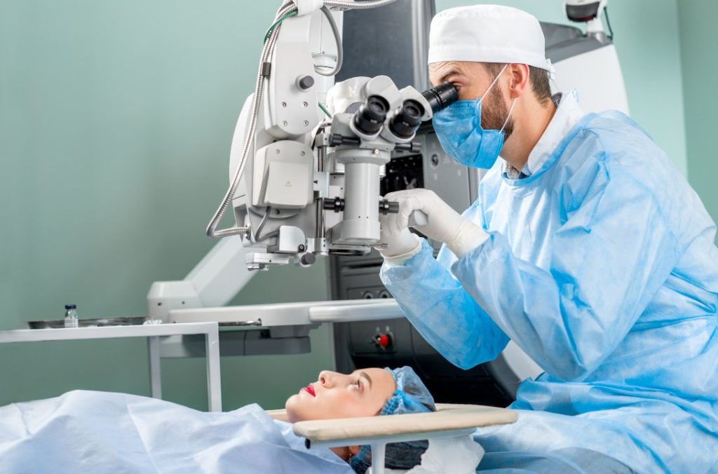
Retinal detachment occurs when the light-sensitive retina separates from the back wall of the eye, and requires prompt surgical repair to prevent permanent vision loss. The main techniques are vitrectomy, scleral buckle, and pneumatic retinopexy, sometimes used in combination. Vitrectomy involves removing the vitreous gel and replacing it with a gas bubble or, occasionally, silicone oil, to hold the retina in place while it heals. A scleral buckle is a silicone band placed around the outside of the eye to relieve traction on the retina. Laser or cryotherapy is used to seal retinal tears. If a gas bubble is used, you may need to keep your head in a particular position for several days, and you must not fly or travel to altitude until the gas has fully absorbed, as this can cause a dangerous rise in eye pressure.
20–30 minutes
Hospital – day case
Local anaesthetic (eye drops)
Permanent – lens lasts a lifetime
1-2 Days
1–2 weeks
~2 weeks (vision-dependent)
Within 24–48 hours
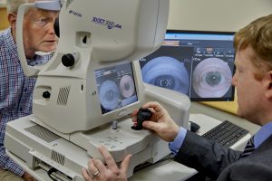
Patients can see Consultant Vitreoretinal Surgeon John Doris in two ways: directly, by booking a consultation themselves, or through a referral from their GP or optometrist/eye specialist. Once seen, Mr. Doris will assess the patient’s condition and recommend an appropriate course of treatment, whether that involves further monitoring, medical management, or surgical intervention.
Anti wrinkle injections can target common signs of ageing and some medical concerns:

A macular hole is a small break in the macula, the central part of the retina responsible for detailed vision, causing blurred or distorted central sight. Surgery involves a vitrectomy to remove the vitreous gel pulling on the macula, followed by peeling of a thin layer of tissue to help the hole close. The eye is then filled with a gas bubble, which flattens the edges of the hole and allows it to heal over several weeks. Vision improves gradually and can take several months to reach its best level. As with any gas-filled eye, flying and travel to altitude are prohibited until the gas has dissolved.
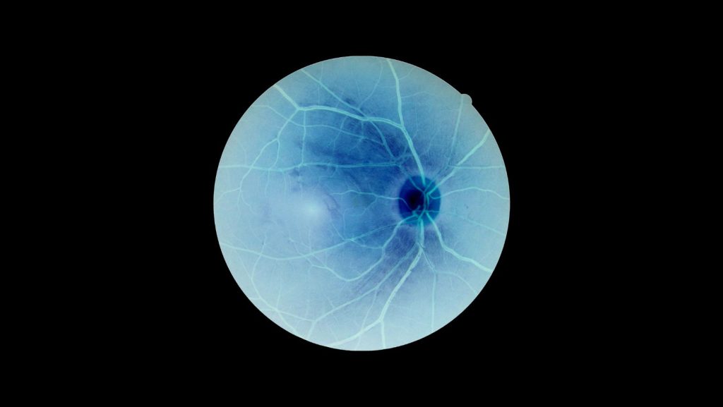
An epiretinal membrane is a thin layer of scar tissue that forms on the surface of the retina, causing blurred or distorted vision, particularly of straight lines. Surgery is usually recommended when symptoms significantly affect daily activities. It involves a vitrectomy to remove the vitreous gel, followed by careful peeling of the membrane from the retinal surface using fine instruments. Most patients do not require a gas bubble, though one is sometimes used. Vision improves gradually over weeks to months, and while distortion often lessens, it may not disappear completely. The main aims of surgery are to stop deterioration and improve, rather than fully restore, vision.
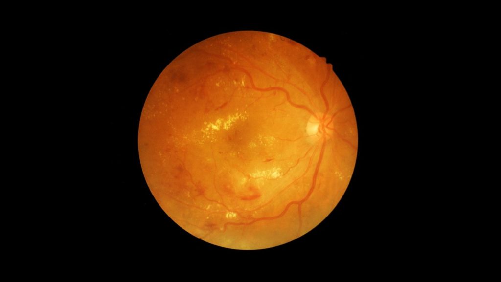
Diabetic eye disease can cause bleeding into the vitreous gel (vitreous haemorrhage), scar tissue formation, and tractional retinal detachment, all of which may require vitrectomy surgery. During the operation, blood and scar tissue are carefully removed from the surface of the retina, relieving traction and allowing the retina to settle back into place. Laser treatment is often applied during surgery to reduce the risk of further abnormal blood vessel growth. A gas bubble, and occasionally silicone oil, may be used to support the retina while it heals. Good control of blood sugar, blood pressure, and cholesterol before and after surgery improves outcomes and reduces the risk of further complications. Recovery can be slower than after other retinal operations, as the underlying diabetic changes in the eye continue to be managed alongside the surgery.
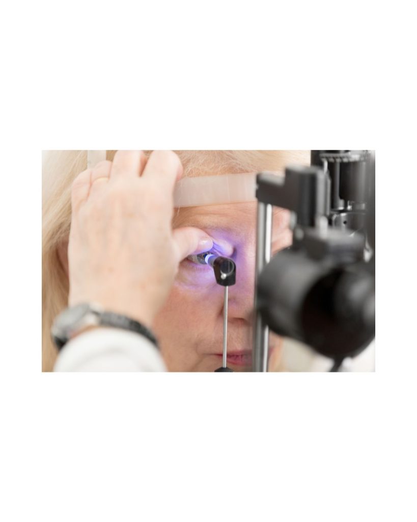
When there is insufficient support to hold an artificial lens in its usual position, for example after complicated cataract surgery or lens dislocation, a lens can be fixed directly to the sclera (the white outer wall of the eye) with fine sutures or a sutureless flanged technique, rather than resting on the eye’s natural support structures. This is known as scleral fixation. The procedure is often combined with a vitrectomy to remove any dislocated lens fragments or vitreous gel that is out of position. Scleral-fixated lenses provide stable, long-term vision correction and avoid reliance on contact lenses or thick glasses. Recovery is generally similar to other vitrectomy procedures, though healing of the scleral wounds may take a little longer, and vision can take some weeks to fully stabilise.
Most retinal surgery can be performed under local anaesthetic, using numbing drops or an injection around the eye. You will be awake but should feel no pain, only occasional pressure or movement. General anaesthetic, where you are fully asleep, may be recommended for longer or more complex operations, for children, for anxious patients, or where someone cannot lie flat or still for the required time. Your surgeon and anaesthetist will discuss the most suitable option for you, taking into account the complexity of surgery, your general health, and your own preference.




Your Journey to Trusted Care Starts Here.
No rush, no pressure – just honest medical advice.

Posted onTrustindex verifies that the original source of the review is Google. The service i received today was exceptional. Dr.Brady was fantastic at explaining, she went above and beyond my expectations.Dr.Brady was extremely kind to me and my farther. As she went into good detail explaing my eyes aswell as farthers. Using great technical terms. Overall Dr. Brady was fantastic.Posted onTrustindex verifies that the original source of the review is Google. Dr. Janice is very welcoming, friendly and professional. She answered all of my questions patiently and with detailed information. I appreciated that she used medical terms during the consultation and then explained the terminology in a simple manner I could comprehend and I am grateful she kindly also used images to further describe various aspects of ophthalmology. I would highly recommend her services and am happy that Dr. Janice was recommended to me by my optician.Posted onTrustindex verifies that the original source of the review is Google. Mr Doris is a fantastic surgeon, totally recommendPosted onTrustindex verifies that the original source of the review is Google. Dr. Janice was patient and thorough, put me at ease and explained every step and every treatment option. Was put at ease by her kind thoughtfulness.Posted onTrustindex verifies that the original source of the review is Google. We’ve had the pleasure of working closely with Eye and Face Clinic Waterford through 2Cubed, and it’s clear why they have such an outstanding reputation. The team there are not only experts in their field but also bring a genuine care and professionalism to every patient interaction. Their approach is thorough, compassionate, and always centred around the patient’s needs. Whether it’s advanced eye care or specialist facial treatments, they combine the very latest techniques with a personal touch that immediately puts people at ease. From our experience, they run their clinic with the same level of dedication and excellence that they bring to their patients – efficient, forward-thinking, and always striving for the best possible results.Posted onTrustindex verifies that the original source of the review is Google. Really enjoyed my first visit to the clinic warm and welcoming reception Gillian is a lovely lady and very passionate about her profession explained treatments that I was interested in can't wait to try the skin pen Katharine CheastyPosted onTrustindex verifies that the original source of the review is Google. Very professional and really helpful would highly recommendPosted onTrustindex verifies that the original source of the review is Google. unfortunately website doesn't workPosted onTrustindex verifies that the original source of the review is Google. The Skin Consultation was excellent, I learned so much about my skin and what products do and don’t work including ingredients to watch out for. I have a totally new skincare regime and already notice a difference after only a couple of weeks. Would definitely recommend for anyone struggling with managing skincare concerns.







Cataract surgery is performed under local anaesthetic eye drops, making it essentially painless. You will be awake throughout and may notice light and movement, but should feel no discomfort. Some mild grittiness or a foreign-body sensation afterwards is normal and usually settles within a day or two.
Most patients notice improvement within 24–48 hours. Vision continues to stabilise over four to six weeks. Some flickering, haloes around lights, or mild haziness in the early weeks is normal and usually settles completely.
This depends on the lens implant chosen. Normally we implant a lens which gives good distance vision but reading glasses will still be required. Your surgeon will advise at your assessment.
In the vast majority of cases the two eyes are operated on separately, typically one to four weeks apart. This allows the first eye to heal and vision to be assessed before proceeding with the second. Your surgeon will advise on timing.
Serious complications are rare. Risks include infection, posterior capsule rupture, retinal detachment, and persistent inflammation. Posterior capsule opacification (a ‘secondary cataract’) can occur months or years later and is easily treated with a quick laser procedure in clinic. Your surgeon will discuss all risks at your assessment.
Intraocular lenses are designed to last a lifetime. Unlike the natural lens, they cannot develop a cataract and do not need replacement or maintenance.
If you experience sudden loss of vision, increasing pain, significant redness, or discharge from the eye, contact our rooms immediately or attend the nearest eye emergency department if outside office hours. Do not wait for your next scheduled review.
You are welcome to contact our rooms directly. Many patients are referred by their GP or optician, but a referral is not always required. If you have been told you may have a cataract, or are experiencing visual difficulty, we would be happy to see you.