
> Eye Care > Eye Conditions > Blind Spot
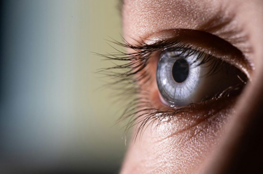
Central vision loss often results from Age related macular degeneration or alternatively a macular hole—both benefit from prompt specialist evaluation.
A macular hole is a small hole in the central part of the retina which can lead to distortion and blurring of central vision. They can be detected by a scan of the back of the eye.
As a hole gets bigger, patients may notice distortion and blurring of vision. The cause of a macular hole is not always clear, but on some occasions may be related to a shrinkage of the jelly filling the posterior part of the eye (the vitreous) which then pulls on the macula (vitreo-macular traction).
Vitrectomy surgery is the main treatment offered and involves the removal of the vitreous gel from the eye, which is then replaced by a bubble of inert gas allowing the hole to close.
As we get older, the macula (the central part of the retina responsible for detailed vision) can begin to break down, causing blurred vision. The macula allows us to see fine detail for activities such as reading, recognising faces, watching television and driving. It also helps us to see colour.
There are two main types of AMD: dry and wet. The extent of the degeneration may be different in each eye and may not progress at the same rate. It is very important to seek early examination and treatment if there is sudden distortion or reduction in vision. There are now some very effective drugs available which can be injected into the eye to stabilise and in some cases improve vision.
This procedure involves removing the vitreous gel (that has caused the retinal tear) from inside the eye. Then to seal the tear the surgeon uses either laser or a freezing probe to make a scar around the tear. A gas or silicone oil bubble is then inserted into the eye to support the retina while it heals. A gas bubble slowly absorbs over 2 to 8 weeks but a silicone oil bubble will need a small operation to remove it at a later date. Your vision will be very blurred initially due to the presence of the gas or oil bubble.
Most retinal tears are small and can be treated using laser or cryotherapy (freezing procedure). This seals the tear and reduces the risk of a retinal detachment.
Retinal detachment surgery can be performed under local anaesthetic or general anaesthetic. Under local anaesthetic you will be awake but you will not feel any discomfort as the eye will be numbed with an injection. You will not see the operation and the other eye will be covered. If a general anaesthetic is chosen then you will be fully asleep. The decision as to which type of anaesthesia is most suitable will be made following a discussion between you and your surgeon.
If you have been given any posturing instructions then these should be followed. You can bath or shower, but avoid splashing water near the eye. Generally you may do anything with which you are comfortable. Most people choose not to drive over the first few weeks. You must not fly until the gas bubble has gone and you must inform the anaesthetist if you require a general anaesthetic for any operation while there is gas in your eye.
Most retinal tears are small and can be treated using laser or cryotherapy (freezing procedure). This seals the tear and reduces the risk of a retinal detachment.
Your Journey to Trusted Care Starts Here.
No rush, no pressure – just honest medical advice.
Posted onTrustindex verifies that the original source of the review is Google. The service i received today was exceptional. Dr.Brady was fantastic at explaining, she went above and beyond my expectations.Dr.Brady was extremely kind to me and my farther. As she went into good detail explaing my eyes aswell as farthers. Using great technical terms. Overall Dr. Brady was fantastic.Posted onTrustindex verifies that the original source of the review is Google. Dr. Janice is very welcoming, friendly and professional. She answered all of my questions patiently and with detailed information. I appreciated that she used medical terms during the consultation and then explained the terminology in a simple manner I could comprehend and I am grateful she kindly also used images to further describe various aspects of ophthalmology. I would highly recommend her services and am happy that Dr. Janice was recommended to me by my optician.Posted onTrustindex verifies that the original source of the review is Google. Mr Doris is a fantastic surgeon, totally recommendPosted onTrustindex verifies that the original source of the review is Google. Dr. Janice was patient and thorough, put me at ease and explained every step and every treatment option. Was put at ease by her kind thoughtfulness.Posted onTrustindex verifies that the original source of the review is Google. We’ve had the pleasure of working closely with Eye and Face Clinic Waterford through 2Cubed, and it’s clear why they have such an outstanding reputation. The team there are not only experts in their field but also bring a genuine care and professionalism to every patient interaction. Their approach is thorough, compassionate, and always centred around the patient’s needs. Whether it’s advanced eye care or specialist facial treatments, they combine the very latest techniques with a personal touch that immediately puts people at ease. From our experience, they run their clinic with the same level of dedication and excellence that they bring to their patients – efficient, forward-thinking, and always striving for the best possible results.Posted onTrustindex verifies that the original source of the review is Google. Really enjoyed my first visit to the clinic warm and welcoming reception Gillian is a lovely lady and very passionate about her profession explained treatments that I was interested in can't wait to try the skin pen Katharine CheastyPosted onTrustindex verifies that the original source of the review is Google. Very professional and really helpful would highly recommendPosted onTrustindex verifies that the original source of the review is Google. unfortunately website doesn't workPosted onTrustindex verifies that the original source of the review is Google. The Skin Consultation was excellent, I learned so much about my skin and what products do and don’t work including ingredients to watch out for. I have a totally new skincare regime and already notice a difference after only a couple of weeks. Would definitely recommend for anyone struggling with managing skincare concerns.